Main results obtained in 2011
The first work package was dedicated to the identification of the system requirements, design of the architecture and specification of the sensor parameters. The total power detection radiometer bloc diagram includes a low-noise amplifier, connected to the antenna. The noise of the low noise amplifier is modeled using the noise temperature TN. The output signal is detected by a square law detection Schottky diode (small signal, x2) and then integrated with a integration time τ, to obtain the root mean square (VOUT) output signal, proportional to the input noise signals. In order to obtain a good image quality in low contrast conditions (inside buildings) it is necessary that the sensor is able to detect temperature differences of 0.5-1 K.
A bibliographic study was performed, and the main issues which will be addressed during the project were identified from literature. A initial parameter database from the electromagnetic area, for different materials, was created. The electromagnetic simulation and modeling techniques were reviewed.
Examples of the variation of the relative permittivity, as a function of frequency, for different biological materials (blood, fat, dry skin), which are important in medical imaging applications, are shown in Fig. 1. The skin depth, a very important parameter for medical imaging, is also presented.
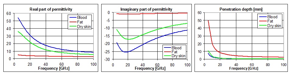
Fig.1 Dielectric properties up to 100 GHz for three biological materials: blood, fat and dry skin