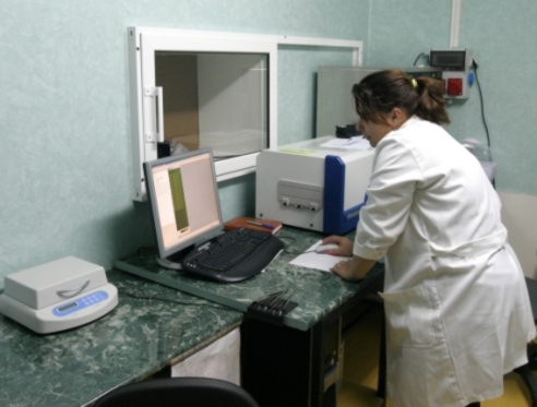
Microarray scanner - GeneTAC UC4
Technical characteristics:
Scanner microarray (GeneTAC UC4) is used for reading the chips, for DNA detecting and deposition → it offers high resolution scanning across the entire surface of standard microarray substrates The system has two-color lasers - green (532nm) and red (635nm) - coupled with high performance optics optimized to maximize collection of fluorescence signal while minimizing the damage caused by photobleaching. The scanner includes: → hardware; → powerful and easy-to-use microarray analysis software for fast and reliable imaging, collection and storage of very large data sets and consolidates these data with experimental information. The entire 1 in. x 3 in. array surface can be imaged at five micron resolution for accurate quantitation of high density arrays. Or a sub area can be scanned at up to single-micron pixel resolution.
- Resolution: from 1 μm/pixel;
- Resolution for a standard microscope slide: 5 μm/pixel
- Microarray Scanner includes also a workstation with powerful software that automates the identification and quantification of microarray data. |
|
| |
Description
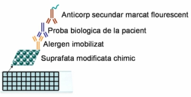
Protein microarray - Depending on the field of application, protein microarrays can be classified into two categories:
(1) Arrays for proteomics or focused protein profiling. This category can be subdivided by array format into forward- and reverse-phase protein microarrays. The difference between the two refers to the way the sample is applied. On forward-phase protein arrays, the sample is incubated on the array so that different analytes can be detected simultaneously. Examples include antibody microarrays that are used for the identification and
quantification of target proteins. On reverse-phase arrays different samples that are immobilised on a chip, in a single step, a large collection of probes can be screened for the presence or absence of one distinct target protein
Protein microarray is used to identify protein-protein interactions, to identify the substrates of protein kinases, or to identify the targets of biologically active small molecules.
(2) arrays for functional studies. Low and high density arrays of proteins, peptides and small molecules could be
used to study the binding of DNA, RNA, small chemical ligands and proteins to their immobilised binding partners.
DNA microarray
A typical microarray experiment compares gene expression levels in two different physiological conditions, for instance one can compare the same bacterial strain grown with or without antibiotics. In this case, nucleic acids of both samples are labeled with distinct fluorescent dyes, most popular dyes are Cy-3 (green) and Cy-5 (red).
****************************************************************** |
Applications:
Protein Arrays
- study tens of thousands of proteins in as short a time as possible;
- producing high density protein arrays on specialized slides;
- automated hybridization and imaging of cDNA and oligonucleotides now allow the consistent high throughput study of antibody/antigen interactions;
Protein Assays
- immuno-assays, protein-protein interaction assays, enzyme assays;
Application procedure
- For each new application it is necessary to be specified the material that should be printed and the spot configuration in order to elaborate the software program;
- A preliminary accept is necessary to avoid the contamination risk;
****************************************************************** |
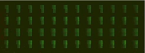
Results:
- In microarray area results are related to functionalization of different surfaces (glass and silicon wafer) in order to immobilized DNA and Proteins used in medical diagnosis. We have optimised the process parameters in order to obtain a uniform hydrophilic/hydrophobic surface, high signal intensity and low background, spot with well defined morphology, and high efficiency with lower cost of slides and chemicals.
-
Enzyme-based sensors designed for electrical measurements of several toxins. The sensors are made of interdigital electrodes built on silicon substrate. Biomaterials are deposited on the electrode surface and are characterized using the NANOBIOLAB equipments.
|
|
Partneship:
-
Multialergen biochip realised by microarray technology, National Project, Program: Parteneriate.Main Partners: University of Bucharest, Faculty of Chemistry, Diagnostic SA (DDS), Genetic Lab.
******************************************************************
|
| Application scientist: phys. Monica Simion, monica.simion@imt.ro |
|
HOME |
 |
Last update: March 06, 2012 |
|