|
Scanning Near-field Optical Microscope (SNOM)
Technical characteristics:
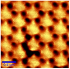
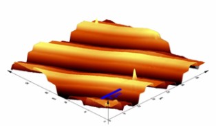
The Alpha300 S System is a Scanning Near-field Optical Microscope (SNOM) that combining the characterization methods of SNOM, Confocal Microscopy (CM) and Atomic Force Microscopy (AFM) in a single equipment. The Alpha300 S uses patented micro-fabricated SNOM cantilever sensors (aperture size typically 100 nm) for optical microscopy with spatial resolution below the diffraction limit (optical resolution of 50 – 100 nm).
Operating modes:
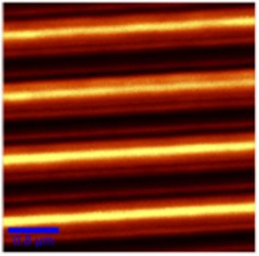
Near field optical microscopy: transmission, reflection, collection, fluorescence
Confocal microscopy: transmission, reflection, fluorescence, can be upgraded with a Raman spectrometer;
Atomic force microscopy: contact and AC – Mode. |
|
Description:
The flexibility of this equipment and its operation modes allows a large variety of applications in nanotechnology and nanosciences :
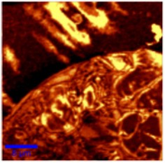
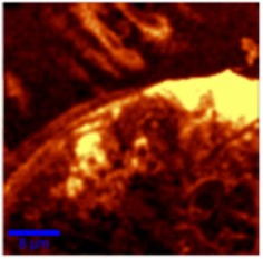
It allows the optical characterization (in both transmission and reflection mode) of various samples (nanostructures, biological samples, polymers) with a resolution of 50-90 nm in visible spectral range with the possibility of extension in the infrared spectral range.
Working in the collection or photon scanning tunneling microscope (PSTM) mode the alpha 300S SNOM allows the imaging of propagating optical field in various metalic and dielectric waveguides providing a powerful method to caracterize and investigate nanophotonics and nanoplasmonic structures and devices.
Also the AFM module working in both contact and alternative contact modes (with posibility of extension to magnetic force measurements and pulsed force mode) allows the topografical and chemical characterization of various surfaces and nanostructures. |
Applications:
- imaging the optical properties of a sample with resolution below the
diffraction limit with applications in nanotechnology, nanophotonics, nanooptics and plasmonics;
- Materials research and polymers
- Single molecule detection;
- Life sciences;
- Fluorescence characterizations
**********************
|
| Results: |
|
Partneship:
- European Centre of Excellence in Microwave, Millimetre Wave and Optical Devices, based on Micro-Electro-Mechanical Systems for Advanced Communication Systems and Sensors - MIMOMEMS (FP7-Capacities) 2008-2010;
- Flexible Patterning of Complex Micro Optical Structures using Adaptive Embossing Technology FLEXPAET (IP- FP7/NMP) 2008-201;
|
| |
| Application scientist: Dr. (phys.) Cristian Kusko, cristian.kusko@imt.ro |
|
HOME |
 |
Last update: March 5, 2012 |
|